Wie kann sich ein Schleimpilz ohne Muskeln fortbewegen?
Ein Schleimpilz hat zwar keine Muskeln wie wir Menschen, doch er besitzt eine Art Prototyp von ihnen. Im Inneren der Plasmodien befindet sich ein feines Zytoskelett, welches aus Aktinfilamenten und dem Protein Myosin aufgebaut ist, und dem Schleimpilz eine Fortbewegung ermöglicht.
How can slime molds move without muscles?
Even though slime molds don't have muscles like we humans do, they possess some kind of a prototype of muscles. Inside a Plasmodium you can find a delicate cytoskeleton, which consists of actin filaments and the protein myosin and allows the slime mold to move.
Zytoskelett, Aktinfilament und Myosin - Was ist das?
Ein Zytoskelett ist im Prinzip eine Art Skelett einer Zelle, jedoch ist dieses nicht aus Knochen aufgebaut, sondern aus Proteinen. Es dient der Stabilisierung und Ordnung in einer Zelle.
Aktinfilamente sind am Zytoskelett verankert, in etwa so wie bei uns Muskeln an Knochen angewachsen sind. Aktinfilamente sind kleine Fasern (= Filament) aus Aktinproteinen, die aneinanderhängen.
Cytoskeleton, actin filament and myosin - What's all that?
A cytoskeleton basically is a kind of skeleton of a cell, but it's not made of bones but proteins. It serves to stabilize and organize a cell.
Actin filaments are anchored in the cytoskeleton, similar to how our muscles are rooted in our bones. Actin filaments are small fibers (= filament) made of actin proteins, which are linked together.
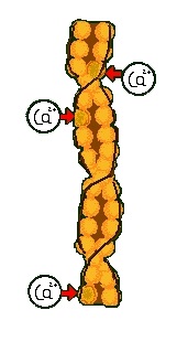
Hier ist so ein Aktinfilament einmal dargestellt. Die einzelnen Kugeln stellen dabei jeweils ein Aktinprotein dar, sie sind schraubenfrömig umeinander gedreht und bilden damit das Filament. Mit im Filament eingebunden sind sogenannte "regulatorische Proteine", die roten Pfeile links zeigen auf welche. Binden Calciumionen an die regulatorischen Proteine, so geben diese Myosinbindestellen frei, sprich Stellen, an die das Protein Myosin binden kann.
Here you can see one of these actin filaments. The individual spheres each represent one actin protein, they are twisted around each other like a screw and thereby form the filament. Enclosed within the filament are so-called "regulatory proteins", the red arrows in the illustration are pointing to them. If calcium ions bind to these regulatory proteins, they release myosin binding sites, thus spots for myosin proteins to bind to.
Ein Myosinmolekül kann man sich dabei in etwa so vorstellen:
A myosin molecule can be imagined like this:
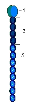
Nummer 1 ist das Köpfchen. Das Köpfchen kann an die Myosinbindestellen des Aktinfilaments binden und spaltet zudem noch ATP. ATP (Adenosintriphosphat) ist ein Molekül mit sehr energiereichen Bindungen. Wird ATP in ADP (Adenosindiphosphat) und Phosphat gespalten, so wird viel Energie frei, die dann andersweitig eingesetzt werden kann. ATP ist somit ein Speicher für chemische Energie.
Nummer 2 ist der bewegliche Hals.
Nummer 3 ist der Schaft.
Viele Myosinmoleküle bilden zusammen ebenfalls Filamente, wobei sich die Schäfte parallel anordnen.
Number 1 is the head. The head can bind to the myosin binding sites of the actin filament and can additionally split ATP. ATP (adrenosine triphosphate) is a molecule with very energy-rich bondings. When ATP is split into ADP (adenosine diphosphate) and phosphate, lots of energy is released, which can then be used otherwise. ATP therefore is a storage for chemical energy.
Number 2 is the movable neck.
Number 3 is the shaft.
Many myosin molecules together also form filaments in which the shafts position themselves parallel to each other.
Insgesamt kann man sich die Anordnung von Myosin- und Aktinfilamenten dann so vorstellen:
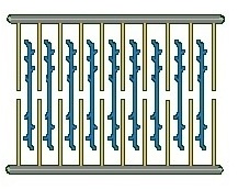
Alltogether, the arrangement of myosin and actin filaments can be imagined like this:
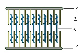
Nummer 1 sind die "Z-Streifen", also die Teile vom Zytosklett, wo die Aktinfilamente verankert sind.
Nummer 2 sind die Aktinfilamente.
Nummer 3 sind die Myosinfilamente. Die Schäfte mehrerer Myosinmoleküle sind in einer solchen blauen Linie parallel angeordnet, die Köpfchen dabei gewinkelt abgestreckt.
1, 2 und 3 bilden zusammen das "Sarkomer".
Number 1 are the "Z-stripes", parts of the cytoskeleton in which the actin filaments are anchored.
Number 2 are the actin filaments.
Number 3 are the myosin filaments. The shafts of multiple myosin molecules are arranged parallel to each other in such a blue line, the heads are bent outwards in an angle.
1, 2 and 3 together form the "sarcomere".
So funktioniert nun die Kontraktion:
Kontrahiert nun ein Muskel (beim Menschen wie auch hier beim Schleimpilz, der zwar keinen Muskel hat, aber eben doch diese Filamentstrukturen), passiert im Einzelnen Folgendes:
Ein Aktionspotential (die Reaktion auf einen überschwelligen Reiz, der die Umpolarisation der Membran verursacht, welche im Ruhezustand innen negativ und außen positiv ist (Ruhepotential, Spannungsunterschied von 70mV)) löst die Ausschüttung von Calciumionen aus, welche sich im Endoplasmatischen Retikulum befinden (dies gilt jetzt vor allem für höhere Lebewesen, für Schleimpilze dürfte dies aber in vereinfachter Form auch zutreffen).
Die Calciumionen binden nun an die regulatorischen Proteine in den Aktinfilamenten. Die Bindungsstellen für Myosin werden freigegeben und Myosinköpfchen heften sich sogleich daran, sie passen hinein wie der Schlüssel in sein Schloss. Das Köpfchen eines Myosinmoleküls besitzt vier Bindungsstellen, welche energetisch unterschiedlich sind. Das Köpfchen rastet zunächst mi der energiereichsten Stelle (1) in die Bindungsstelle am Aktinfilament ein.
And this is how the contraction works:
When a muscle contracts (in a human body as well as in a slime mold, which doesn't have muscles but these same filament structures), this is what happens in detail:
An action potential (the reaction to an above-threshold stimulus, which causes the repolarization of the membrane, which, in its idle state, is negative on the inside and positive on the outside (restingpotential, potential difference of 70mV)) causes the release of calcium ions, which are located in the endoplasmatic reticulum (this is mostly true for higher beings, but probably works the same way, maybe simplified, in slime molds).
The calcium ions now bind to the regulatory proteins in the actin filaments. The binding sites for myosin are uncovered and myosin heads immediatly affix to them. They fit into these binding sites like a key in its lock. The head of a myosin molecule possesses four binding sites, which are energetically different. The head first locks in with the most energetic (1) into the binding site on the actin filament.
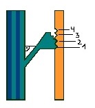
Der Winkel unter dem das Myosinköpfchen in die erste Bindungsstelle einrastet beträgt 90°.
The angle at which the myosin head docks into the first binding site is 90°.
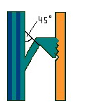
Nach und nach rastet das Köpfchen nun in die energieärmeren Bindungsstellen. Ist das Köpfchen im energieärmsten Bindungsort (4) gebunden, so beträgt der Winkel, unter dem es abgeknickt ist, nur noch 45°.
The head then locks into the less energetic binding sites step by step. Once the head is locked onto the least energetic binding site (4), the angle, to which the myosin head is bent, is only 45°.
Für das Abknicken des Köpfchens ist der bewegliche Teil des Halses notwendig.
The bending of the head would not be possible without the movable neck of the myosin molecule.
Als Folge dieses Anhaftens und Abknickens des Köpfchens, wird das Myosinfilament im Aktinfilament verhakt und verschiebt sich somit näher an die Z-Streifen. Der Abstand zwischen den beiden Z-Streifen wird somit verringert:
Der Muskel ist damit kontrahiert.
As a result of the adhesion and bending of the head, the myosin filament gets caught up in the actin filament and moves closer to the Z-stripes. The distance between the two Z-stripes therefore decreases:
The muscle contracted.
Bindet nun ATP in die katalytische Stelle des Myosinköpchens, so wird dieses gehemmt. Das Köpfchen verändert seine Struktur und kann nicht mehr an das Aktinfilament binden, es löst sich. ADP und Phosphat verlassen das Köpfchen, welches wieder in seine Ausgangslage zurückklappt und erneut an das Aktinfilament binden kann. Somit zieht sich das Myosinfilament immer näher zu den Z-Streifen, der Muskel kontrahiert weiter.
If ATP now binds to the catalytic site of the myosin head, it gets obstructed. The head changes its structure and can't continue binding to the actin filament, it detaches. ADP and phosphate leave the head, which folds back into its initial state and can then begin to bind to the actin filament again. Therefore, the myosin filament keeps pulling itself closer and closer to the Z-stripes, the muscle contracts further.
Die Muskelentspannung
Lässt die Erregung wieder nach, so werden keine weiteren Calciumionen freigesetzt und die regulatorischen Proteine verdecken wieder die Myosinbindungsstellen. Restliche Calciumionen werden durch Ionenpumpen in das Endoplasmatische Retikulum zurückgepumpt, der Muskel entspannt.
Nach diesem Prinzip enstehen auch die Kontraktionen in einem Schleimpilz. Es kontrahiert zwar kein ganzer Muskel, dafür aber die Plasmamembran und das Zellplasma.
Muscle relaxation
Once the excitation ceases, no more calcium ions are released and the regulatory proteins cover the myosin binding sites. Left over calcium ions are pumped back to the endoplasmatic reticulum via ion pumps, the muscle relaxes.
This is also how the contractions in a slime mold work. Even though it doesn't cause a whole muscle to contract, it contracts the plasma membrane and the cytoplasm.
Weitere Eigenschaften der Fortbewegung bei Schleimpilzen:
Mikrotubuli: Zusätzlich verfügt ein Schleimpilz über sogenannte Mikrotubuli, röhrenförmige Proteinfilamente, die ebenfalls für die Bewegung, Stabilisierung und Transport von Vesikeln (kleinen Transportmolekülen mit irgendwelchen Stoffen drinnen) zuständig sind. Solche Transporte entlang von Proteinfilamenten sind lebensnotwendig, da reine Diffusion wegen der Größe der Plasmodien nicht mehr ausreichend wäre.
Plasmaströmungen: Beide Proteinfilamentstrukturen sind zuständig für Plasmaströmungen, die die Fortbewegung ebenfalls fördern. Kontrahieren Plasmamembran und Zellplasma, so fließt das Plasma an Orte geringeren Drucks.
Die Plasmaströmungen in Schleimpilzen liegen mit 1000µm/sec deutlich über denen in Pflanzenzellen (2-78µm/sec).
Zellorganelle: Die Zellorganelle in einem Schleimpilz können sich entlang der Filamente mithilfe von Motorproteinen ebenfalls fortbewegen.
Fazit bei der Beobachtung: Insgesamt sieht man bei genauer Beobachtung am Schleimpilz keine gleichmäßigen Bewegungen, sondern eine Kombination von pulsierenden Rück- und Vorstößen. Vor einer Vorwärtsbewegung zieht sich das Plasmodium zunächst kurz zurück, ehe es nach vorne stößt.
Further properties of a slime mold's locomotion:
Microtubules: Slime molds additionally have so-called microtubules at their disposal, tubular protein filaments which are also responsible for motion, stabilizing and the transport of vesicles (small transport molecules that carry certain substances). Such transports along protein filaments are vital for life, because pure diffusion would not be enough for the size of slime mold plasmodia.
Cyclosis: Both protein filament structures are responsible for cycloses, which support the locomotion. When plasma membrane and cytoplasm contract, the plasma flows to areas with less pressure.
The cyclosis in slime molds happens at a rate of 1000µm/sec, much faster than those in plant cells (2-78µm/sec).
Cell organelles: The cell organelles in a slime mold can also move along the filaments using motor proteins.
Conclusion of the observation: Overall, when observing a slime mold one doesn't see a constant motion, but a combination of pulsing back and forward movements. Before moving forward, a plasmodium first pulls itself backwards shortly before pushing forward.